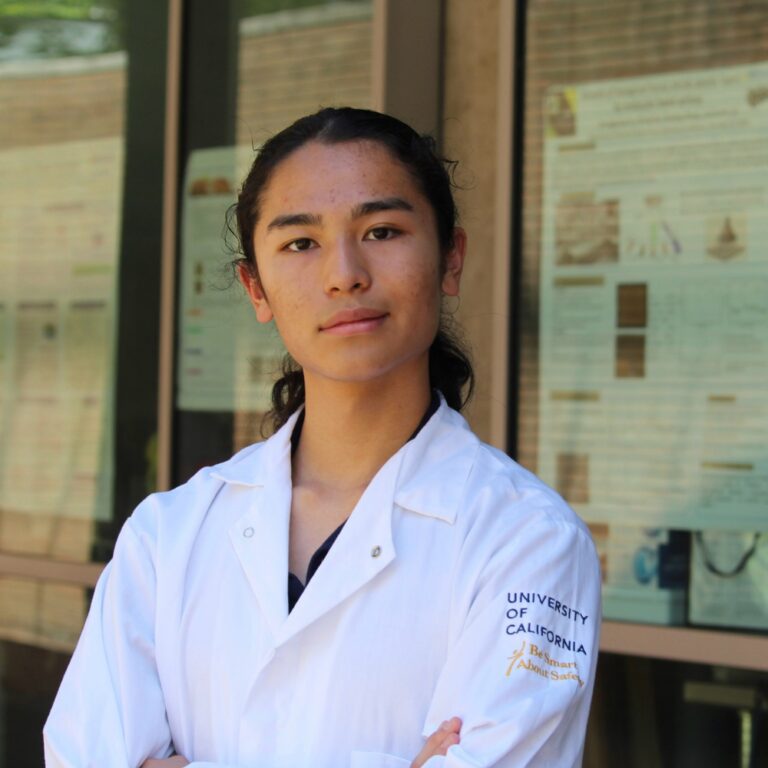
Poster #108, University of California, Riverside
A Multi-View Cross-Referencing Approach to Manual Annotation of 4D Echocardiographic Images for Deep Learning-Based Tricuspid Valve Segmentation
Mentors: Maedeh Makki, MS; Ashley Taepakdee, MS; Zachary Molander, MS; PI: Chung-Hao Lee, PhD
Patient-specific computational models have become valuable tools for understanding heart valve biomechanics and advancing regenerative approaches for congenital and acquired heart diseases. However, the accuracy of these models is often limited by the quality of anatomical data used to define valve geometry. Inaccurate geometric reconstruction may introduce errors in predicted tissue deformation and stress distributions, potentially limiting the efficacy of regenerative scaffolds for stem cell-based valve therapies. Improving annotation precision and geometric fidelity enhances the reliability of downstream biomechanical analysis. This study is part of a larger effort to automate extraction of tricuspid valve geometry with U-Net architecture for integration with patient-specific finite element simulations and mechanically informed regenerative scaffold designs. In conventional methodology, cross-sections of the tricuspid valve are individually annotated along sagittal or coronal planes. While this method produces comparatively rapid results, significant loss of annotation precision may occur in regions tangential to the imaging plane due to perspective-dependent landmark ambiguity. Such geometric inaccuracies may alter reconstructed valve morphology and influence predictions of tissue mechanics, decreasing model reliability. This project introduces a multiview cross-referencing approach to mitigate these limitations and improve spatial accuracy of valve reconstruction. Following the annotation of four-dimensional echocardiography images, the resulting landmark point clouds are propagated across intermediate time frames using end-to-end spatiotemporal tracking algorithms such as MyoTracker to reduce tracking drift and increase robustness to image artifacts. This approach may improve extraction of anatomical datasets for predictive modeling of patient-specific tricuspid valve geometry and functional biomechanics. Future research may apply finite element analyses to define patient-specific mechanical environments that support the development of valvular endothelial and valvular interstitial cell-seeded scaffolds.